

Breast cancer is very heterogeneous. In other words, there are different types of breast cancer, depending on:
- Tumour location
- Grade
- Migratory state of the cancer cells
- Stage
- Molecular subtype
- Gene mutation
The tests listed in the “Diagnostic Tests” section determine the type and stage of the cancer. The abovementioned cancer factors can have approximately 150 combinations, each with its own risk of progressing, metastasizing and recurrence. Determining the type of cancer is key to making the best possible choice of treatment.
Type Of Breast cancer
Cancer characterization takes into account the location of the cancer cells:
- Ductal carcinoma: the cancer cells are in the breast’s lactiferous ducts. This type represents 85% of breast cancers.
- Lobular carcinoma: the cancer cells are present in the lobules. This type represents 10-15% of breast cancers.
Rate types of Breast cancer
- Inflammatory breast cancer (IBC)
- Paget’s disease of the breast
- Diffuse large B-cell lymphoma (non-Hodgkin’s lymphoma type)
- Soft-tissue sarcoma
- Melanoma
- Metaplastic tumours
- Adenoid cystic carcinoma
- Phyllode tumour
- Carcinosarcoma
- Basal breast cancer
The tissue affected can be seen [3] via ultrasound or mammography.
Inflammatory Breast cancer (IBC)
Rarer types of breast cancer include inflammatory breast cancer (IBC) and Paget’s disease. Unlike most other breast cancers, which give rise to one or more separate solid tumours, inflammatory breast cancer tends to form “sheets” or “nests.”
The cancer cells then block the lymphatic system locally, causing these specific signs and symptoms of IBC:
- Redness over more than a third of the breast, with or without heat;
- Persistent itching;
- Increased breast volume;
- Swelling or hardening of the skin, which may take on the look of an orange peel;
- Nipple retraction (turned inwards).
There may be one or more of these signs, and this form of cancer can sometimes be painful.
IBC can be difficult to detect since many of its symptoms are similar to those of other, more common health issues, like mastitis or breast infection which both cause inflammation of the breast. This often leads to diagnosis in Stage III or IV. This form of cancer can be particularly aggressive and progress rapidly. Do not wait to consult a healthcare professional if the signs of inflammation do not diminish after 7 to 10 days of treatment.
Learn more about inflammatory breast cancer:
Canadian Breast Cancer Network
Cancer Foundation (Belgium)
American Cancer Society
Moose & Doc Breast Cancer
Learn more about Paget’s disease:
Quebec Breast Cancer Screening Program (Capitale-Nationale region) (in French only)
Grade
An important characteristic of cancer is its grade, which considers the cells’ differentiation, growth rate, structural abnormalities and death rate (tumour necrosis). The grade is the cancer’s degree of malignancy.
Differentiation
The morphological characteristics of cancer cells are different from those of normal cells. A cancer cell that looks like a healthy cell is said to be “well-differentiated,” while a cell that looks very different is “poorly differentiated” or “undifferentiated” (it loses the characteristics of the original cell). The less differentiated the cell is, the higher the grade, indicating that the cancer is more likely to grow rapidly.
Differentiation is determined by observing the morphology of the cells taken during the biopsy.
Growth rate
To assess the growth rate of the cancer cells, Ki-67 expression levels are measured. This protein increases in quantity as the cells divide. Immunohistochemistry is used to immunostain the cells containing Ki-67 to determine their percentage.
The more positive cells there are, the faster the cancer is progressing.
| GRADE | DESCRIPTION |
|---|---|
| 1 | Slow growth – Low risk of spread |
| In situ tumours | Well-differentiated cancer cells; resemble normal cells |
| Invasive tumours | Small-to-medium cancer-cell nuclei; all have the same shape No evidence of cellular necrosis |
| 2 | Growth and average risk of spread |
| In situ tumours | Moderately differentiated cancer cells |
| Invasive tumours | Small-to-medium cancer-cell nuclei; all have the same shape Some small areas of cellular necrosis |
| 3 | Rapid growth – High risk of spread |
| In situ tumours | Poorly differentiated or undifferentiated cancer cells; abnormal appearance in comparison to healthy cells |
| Invasive tumours | Large cancer-cell nuclei; uneven shape Cellular necrosis |
Migration of cancer cells
- In situ (non-invasive) tumours: The cancer cells do not invade other organs in the body.
- Infiltrating/invasive tumours: The cancer spreads beyond the layer of tissue in which it originally developed. Cancer cells have moved to tissue around the lactation ducts, including the lymph nodes, via the blood vessels or lymphatic ducts.
- Metastatic tumours: Infiltrating cancer cells can travel through the body and attach to other organs.
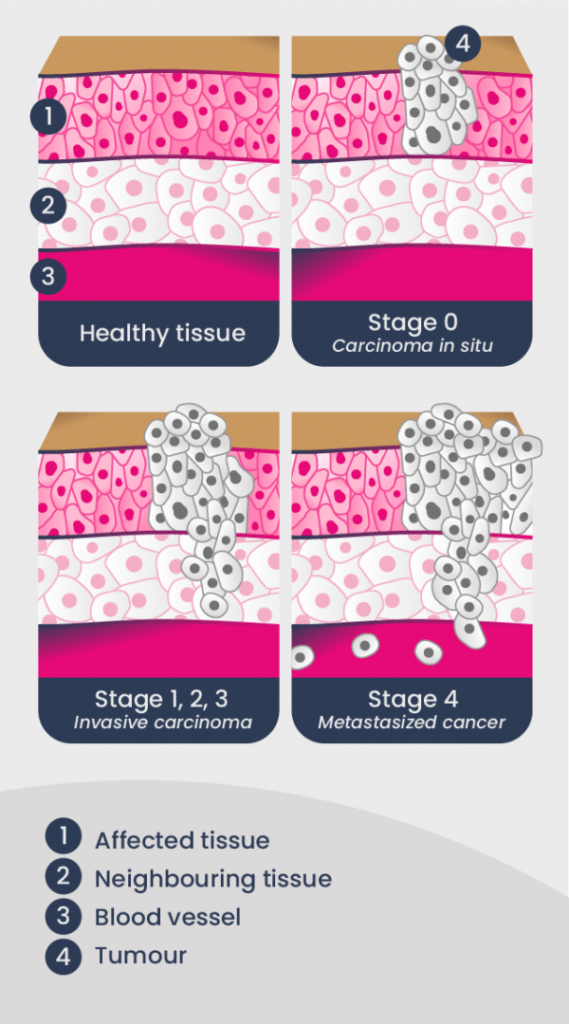
Cancer Stages
The stage of cancer is measured by how far the cancer cells have spread. Depending on its stage, the cancer is said to be early or advanced.
The TNM system (for “tumour, node, metastasis”) is used to determine the stage of the cancer. This evaluation takes into account the size of the tumour, the number of lymph nodes containing cancer cells and the presence of cancer in other organs.
Tumour size is measured by ultrasound or mammography, while lymph nodes are examined by clinical examination, sentinel node biopsy or axillary lymph node dissection. Additional tests (ultrasound of the liver, x-ray of the lungs, bone scintillography and blood tests to assess the condition of the organs) are used to test for metastasis.
| STAGE | CARACTERISTICS |
|---|---|
| 0 | The cancer cells are located only within the membrane of a lactiferous duct (i.e., ductal carcinoma in situ) or lobule (i.e., lobular carcinoma in situ). |
| 1 | The cancerous tumour is 2 centimetres or less in size and has not spread beyond the breast. |
| 2 | The cancerous tumour is 2-5 centimetres or less in size, has spread to nearby lymph nodes, or both. |
| 3 | The cancer has spread to the lymph nodes and possibly to nearby tissues, such as muscles or skin. |
| 4 | The cancer has spread to other parts of the body. |
Molecular Subtypes
Disruption of the expression of ER- and PR-hormone receptors (i.e., the amount of protein they produce) and/or HER2-receptor is often involved in the development of breast cancer.
It is important to determine whether these receptors are overexpressed (i.e., present in greater-than-usual amounts) in cancer cells, since the level of their expression determines what treatment will be given.
To evaluate the expression of hormone and HER2 receptors, tests are done on biopsied cancer cells.
| Receptors | |
| ER- and PR-hormone | Her2 |
| Prevalence | |
| 83% of breast cancers | 17% of breast cancers |
| Traitements | |
| Hormone therapy | HER2-targeted therapy (e.g. Trastuzumab) |
| Diagnostic test | |
| Immunohistochemistry | Immunohistochemistry FISH |
Immunohistochemistry (IHC) is used to stain the cells expressing the receptors. Fluorescence in situ hybridization (FISH) measures the number of copies of the HER2 gene in cancer cells.
Triple-negative Breast cancer
When the cancer does not express any of these receptors, it is called triple-negative breast cancer. The treatment of this type of cancer will differ from that of cancers that overexpress ER- and PR-hormone or HER2 receptors.
- 10-15% of breast cancers are triple-negative.
- It includes several types of cancers, such as basal (70% of triple-negative cancers) and cancers with BRCA1 and BRCA2 mutations (20%). It also groups cancers with androgen-receptor expression and mesenchymal cancer.
- It is a usually an aggressive cancer, and more often affects younger or obese women.
- It does not respond to hormone therapy or HER2-targeted therapy. Chemotherapy is usually used for this type of cancer. Several treatments are under investigation for this type of cancer.
Mutations in Key genes
The doctor may order a genetic analysis of the tumour, to check for the mutations or overexpression of genes known to be defective in breast cancer. Defects in the DNA sequence indicate a more aggressive cancer with a higher risk of recurrence. In these cases, chemotherapy is often administered to eliminate the cancer more effectively.
The Oncotype DX genetic test analyzes the expression or activation of 21 genes, and reports results with a score of 0 to 100. The lower the number, the lower the risk of recurrence and the less likely chemotherapy will be recommended.